Having a loved one in a coma or unconscious can be a tough, nerve wracking experience for all involved. This type of situation is also stressful for the doctor because it is often very difficult to predict whether the patient will remain in a permanent vegetative state, or if the patient will one day recover and return to a normal life.
The pressure only increases when the patients’ families are trying to decide whether or not to remove the individual from life support and they are looking to the doctor for their deciding opinion; an opinion and prediction which is always difficult.
Fortunately, technology is improving and scientists and doctors may have found a way to make a much more accurate diagnosis.
Table of Contents
Background and Terminology
There are two different states that are experienced by people who are considered to be in a coma. The first is referred to as a “Minimally Conscious State,” or MCS. In this state, the patient has the ability to respond to stimuli and has the potential to fully regain consciousness.
However, people who are in a permanent vegetative state (VS) are unable to respond to stimuli in any way and is very unlikely to ever recover. And since VS and MCS are different from being brain dead, meaning that there is still some brain function, it can be very difficult for a doctor to distinguish between the two states.
A Study in Brain Scans
A recent study that uses brain scans to diagnose whether a patient is in MCS or VS, was reported by CNN and revealed the following:
The Lancet study authors looked at the potential of functional brain imaging to help doctors predict whether patients will regain consciousness. The study was conducted at a hospital in Belgium. Doctors tracked 81 patients who had been diagnosed as being in a minimally conscious state and 41 who’d been diagnosed as being in a vegetative state.
Up to 40% of patients are misdiagnosed in these situations, researchers wrote in the accompanying editorial. Four patients who were conscious but unresponsive were used as a control group. Doctors looked at functional MRI and PET scans for all patients.
The Results
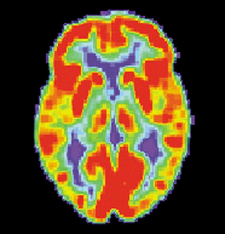
The researchers who conducted the study found that PET imaging was significantly more accurate that MRIs in distinguishing the likelihood of a patient recovering. Over the course of the year in which this study was conducted, PET scans showed a 74% accuracy rate, compared to an accuracy rate of 56% by MRIs, following the predictions by prior neurobiological research.
The reason for the difference in accuracy is that PET scans take measurements of a patient’s brain’s metabolic activity, rather than simply looking at the brain’s structure, which is what an MRI does.
The Takeaway
CNN reported on the researchers’ conclusions saying:
PET scans may eventually help doctors diagnose patients with serious brain injuries, lead study author Steven Laureys says, but it’s not ready for widespread use yet. The study was done in a special unit with trained researchers. Using PET as a tool for diagnosis of brain-injured patients too soon could do more harm than good – leading to more misdiagnoses instead of less.
Sleigh and Warnaby commented on the findings saying, “Functional brain imaging is expensive and technically challenging, but it will almost certainly become cheaper and easier. In the future, we will probably look back in amazement at how we were ever able to practice without it.”
What does this Mean for You?
This means that if you or someone you know is involved in an accident and is in a coma, you may want to ask about this research and see if your doctor can use the findings to more accurately diagnose the patient. Losing a loved one is never an easy experience, but to be unsure of whether or not you are making the right decision can stay with you for life. At Good Guys Injury Law, we want to make sure that you have all the resources necessary to better your life and the lives of those you love.
Photo courtesy of US National Institute on Aging, Alzheimer’s Disease Education and Referral Center

